

An ECG (Electrocardiogram) is a medical test that records the electrical activity of the heart over a period of time. It is a simple, non-invasive procedure used to monitor the heart’s rhythm, detect heart diseases, and assess the heart’s function. An ECG provides important information about the heart’s electrical system, which controls the timing and coordination of heartbeats.
How ECG Works:
- The heart beats due to electrical impulses that trigger the contraction of heart muscles. These electrical signals are generated by the heart’s natural pacemaker, the sinoatrial (SA) node, and travel through the heart to coordinate its pumping action.
- An ECG records these electrical impulses by attaching electrodes (small, sticky sensors) to the skin on various parts of the body, such as the chest, arms, and legs. The electrodes detect the electrical activity of the heart and transmit this information to an ECG machine, which produces a graphical representation of the heart’s electrical activity.
ECG Waveforms:
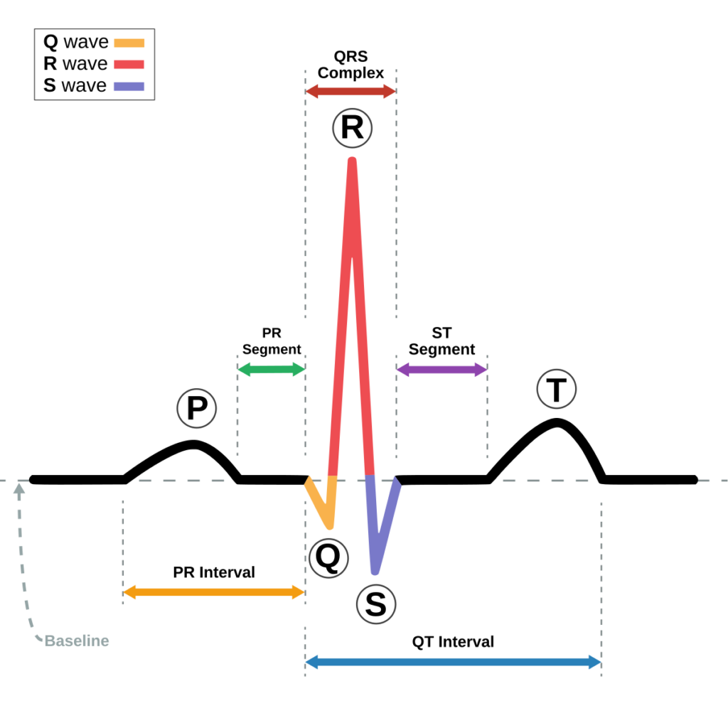
- The ECG machine records the electrical activity as a series of waves or tracings, known as the P wave, QRS complex, and T wave, each corresponding to a specific part of the heart’s electrical cycle:
- P wave: Represents the electrical impulse that causes the atria (upper chambers) to contract.
- QRS complex: Represents the electrical impulse that causes the ventricles (lower chambers) to contract.
- T wave: Represents the recovery (repolarization) of the ventricles after contraction.
- The shape, timing, and duration of these waves help doctors analyze the heart’s rhythm, rate, and overall health.
- The ECG machine records the electrical activity as a series of waves or tracings, known as the P wave, QRS complex, and T wave, each corresponding to a specific part of the heart’s electrical cycle:
Types of ECG:
- Resting ECG: The most common type of ECG, taken while the patient is lying down and resting. It provides a baseline measurement of the heart’s electrical activity.
- Stress (Exercise) ECG: Performed while the patient is exercising on a treadmill or stationary bike. It helps assess how the heart responds to physical stress and is commonly used to diagnose heart disease or evaluate heart function in people with chest pain or risk factors.
- Holter Monitor: A portable, continuous 24-48 hour ECG that records the heart’s activity throughout the day. It is useful for detecting irregular heartbeats that may not appear during a standard ECG.
- Event Monitor: Similar to a Holter monitor, but it is worn for a longer period (up to a month), and it records the ECG only when the patient feels symptoms (e.g., palpitations). The patient can activate the device to record the event.
- Ambulatory ECG: A longer-term ECG recording device that is worn outside the hospital for extended periods to monitor the heart’s activity during daily activities.
ECG Leads:
- An ECG test typically uses 12 leads or electrodes to measure electrical activity from different angles, providing a comprehensive view of the heart’s electrical system.
- The leads are placed on the chest and limbs in specific locations, allowing for the detection of electrical activity from various perspectives (e.g., front, back, left, right) of the heart.
Purpose of an ECG:
Assess Heart Rhythm and Rate:
- An ECG is commonly used to assess the heart’s rhythm and rate. For example, it can detect abnormal rhythms, such as arrhythmias, which may cause symptoms like dizziness, palpitations, or fainting.
Diagnose Heart Diseases:
- Coronary artery disease (CAD): ECGs can show signs of reduced blood flow to the heart muscle, which may indicate blockages in the coronary arteries.
- Heart attacks: During or after a heart attack, an ECG can reveal abnormalities in the heart’s electrical activity, such as ST-segment elevation, which suggests that part of the heart muscle is not receiving enough oxygen.
- Cardiomyopathy: ECG can help diagnose diseases that affect the heart muscle’s ability to pump blood effectively.
Monitor Heart Health:
- ECGs are often used to monitor the heart’s condition in patients with known heart disease, ensuring that treatment is working effectively and there are no dangerous changes in heart rhythm.
Electrolyte Imbalance and Other Conditions:
- Electrolyte disturbances: Conditions like high or low potassium or calcium levels can affect the heart’s electrical activity, and ECGs can help detect these imbalances.
- Pericarditis: Inflammation of the heart’s outer lining can show up as changes in the ECG, such as widespread ST-segment elevation.
Pre-Surgical Evaluation:
- ECGs are often done before surgery to ensure that the patient’s heart is in a stable condition and to assess any potential risks related to anesthesia or surgery.
Assess Effectiveness of Medications:
- Doctors may use ECGs to monitor the effectiveness of treatments for arrhythmias, heart failure, or other cardiac conditions and to detect side effects of medications that may affect the heart’s electrical activity.
Common Conditions Detected by ECG:
Arrhythmias:
- Abnormal heart rhythms, such as atrial fibrillation (AFib), ventricular tachycardia, or bradycardia, can be detected by ECG. Arrhythmias can cause symptoms like heart palpitations, dizziness, or fainting.
Myocardial Infarction (Heart Attack):
- An ECG can help identify a heart attack (myocardial infarction) by looking for signs of damage to the heart muscle, such as ST-segment elevation or inverted T waves.
Heart Block:
- This occurs when the electrical signals in the heart are delayed or blocked, and it can be identified by abnormal patterns in the P-R interval or QRS complex.
Electrolyte Imbalances:
- Low or high levels of certain electrolytes (like potassium or calcium) can affect the heart’s rhythm and lead to abnormal ECG findings.
Congenital Heart Disorders:
- ECGs can sometimes detect heart defects present at birth that may affect the heart’s electrical conduction system.
How the ECG Test Is Performed:
- Preparation:
- The patient is usually asked to remove clothing from the upper body, as the electrodes need to be attached to the skin.
- A gel or adhesive is sometimes applied to improve electrode contact with the skin.
- Electrode Placement:
- Small, sticky pads (electrodes) are placed on the chest, arms, and legs. For a 12-lead ECG, 10 electrodes are typically used.
- Recording:
- The patient is asked to lie still and breathe normally while the ECG records the heart’s electrical activity for a few minutes.
- Interpretation:
- Once the test is complete, the results are analyzed by a doctor or cardiologist, who looks for any irregularities or signs of heart disease.
Advantages of ECG:
- Non-Invasive: The procedure does not involve any needles or surgery.
- Quick and Painless: ECGs are fast (usually taking about 5–10 minutes) and do not cause discomfort.
- Cost-Effective: ECG is a relatively inexpensive test compared to other imaging tests like CT scans or MRIs.
- Widely Available: ECG machines are available in hospitals, clinics, and even some ambulances, making the test easily accessible.
Limitations of ECG:
- Does Not Show Detailed Heart Anatomy: While an ECG shows the electrical activity of the heart, it doesn’t provide detailed images of the heart’s structure or blood flow. For this, additional tests like echocardiography or angiography may be needed.
- Not Always Conclusive: Some heart conditions may not be detectable by an ECG, especially if the symptoms are intermittent or if the test is done at a time when the abnormality is not occurring.